Thursday, January 31, 2019

Introduction.

Every human being in this world has his own talent, to create  or to say something, that may be usefull to sombody or to many people.,but fear to make a mistake discourage or stop many of us,to share their knowledge to others. And that is a greate mistake,  because nobody about a mistake.and we have to realize that making a mistake for hundread times, is mor better than not to try at all. And a mistake is not a weakness,the weakness is ,not to accept your mistake after its had been discovere by  sombody,who may be your senior or your junior.the father of  of relativity said (Albert einstein), " the brain that creates a problem should not be use to solve that particular  problem.means there is need to apply another brain to solve that particular problem".
 So that is why  i am trying to create this blogin website ,for the purpose of teaching and learning, in the science-related stuff.
as everyone knows, science is a collection of many ideas,theories and hypothesis,which evolve from the ancient time to till- date.in this science turtorial concepts ,we will focus on five main science courses,Wich are;
1). Life science.
2). earth science.
3).Computer science.
4) engineering.

 We will start from the life-related science,which is biology.
biology is a science that deals with life or living thing.which compose of many creatures from one cell (uni-cellular orgernsim) to  insects,birt,fish and human being,whose are (multicellular organisims) .
 And in this  broad amazing course. we will still start from the study of human body, or (human biology),because it is the most important.why?,,because you as a human being ,knowing how your body and your  internal organs work.will make you very easy to maintain them in proper way.
As every organisms,human biological structure started from cell, to tissue,organ,and organs-system.
for good foundation,let us start from the cell ,then to tissue,organ,and the organs-system.
Another advantage of this site is that: everything will explain in simple-english-words, in other to be understand by the even road-site-men.

and for those who want to read these turtorial in hausa language,they can visit my other website,at
www.kimiyyadahausa.blogspot.com
where I  explained everything in our mother's language.(hausa).thanks to everybody.
#For any corrections or ssuggestion, just  let me know in the below box, or econtact me at junaiduumar2233@gmail.coml

Wednesday, January 30, 2019

(1).Science.

Science is simply means;the system ore ability of human being to understand how the world's nature works or behave.
There are a lot of things in this world,which men have been doing or using from the ancient times to till-date. But They do not know the science behind them.and knowing lt,will leads us to better understanding  our physical world & its laws. And This also help and serves as the foundation stone for our technologies to evolve,in  hausa community and around the globe. Reading more on physical,chemical and biological phenomener, of this world,will make our   life very easy.
 So in this turtorial,I will guid me and my fellow brothers and sisters,on how to do this in simple manner,by providing us a science-related lessons, in a simple and understadable words,  in other to be comprehen by the even primary student.and if you are not good in science ,at all or you are not familiar With science-relateted-words,just skip this site and jump to my other website at
www.kimiyyadahausa.blogspot.com where everything explained in hausa language.
god bless all of us.

Monday, January 28, 2019

(2).Cell

Cell .
What is a cell? Cells are the basic building blocks of all living things. The human body is composed of trillions of cells.
It is the cells that provide structure for the body,cells  take in nutrients from food, convert those nutrients into energy, and carry out specialized functions. Cells also contain the body’s hereditary material and can make copies of themselves. Cells have many parts, each with a different function. Some of these parts, called organelles, are specialized structures that perform certain tasks within the cell. Human cells contain the following major parts, .

1). Cytoplasm :

the cytoplasm is made up of a jelly-like fluid (called the cytosol) and other structures that surround the nucleus.

 2).cytoskeleton;

 The cytoskeleton is a network of long fibers that make up the cell’s structural framework. The cytoskeleton has several critical functions, including determining cell shape, participating in cell division, and allowing cells to move. It also provides a track-like system that directs the movement of organelles and other substances within cells.

3).Endoplasmic

reticulum (ER) This organelle helps process molecules created by the cell. The endoplasmic reticulum also transports these molecules to their specific destinations either inside or outside the cell.

4).Golgi apparatus;

 The Golgi apparatus packages molecules processed by the endoplasmic reticulum to be transported out of the cell.

4).Lysosomes;

and peroxisomes These organelles are the recycling center of the cell. They digest foreign bacteria that invade the cell, rid the cell of toxic substances, and recycle worn-out cell components.

5). Mitochondria

 ;Mitochondria are complex organelles that convert energy from food into a form that the cell can use. They have their own genetic material, separate from the DNA in the nucleus, and can make copies of themselves.

6). Nucleus

;The nucleus serves as the cell’s command center, sending directions to the cell to grow, mature, divide, or die. It also houses DNA (deoxyribonucleic acid), the cell’s hereditary material. The nucleus is surrounded by a membrane called the nuclear envelope, which protects the DNA and separates the nucleus from the rest of the cell.

7). Plasma membrane;

The plasma membrane is the outer lining of the cell. It separates the cell from its environment and allows materials to enter and leave the cell.

8). Ribosomes

; Ribosomes are organelles that process the cell’s genetic instructions to create proteins. These organelles can float freely in the cytoplasm or be connected to the endoplasmic

reticulum.for further understanding read it in this website,
www.kimiyyadahausa.blogspot.com

Sunday, January 27, 2019

(3).Tissue

Tissue;
tissue consists of groups of cells with a similar structure working together for a specific function. 
Human body tissue is another way of describing how our cells are grouped together in a highly organized manner according to specific structure and function. These groupings of cells form tissues, which then make up organs and various parts of the body.
 Types of Tissue in Human Body ;)
1).1.Muscl :
Muscle tissue is made up of excitable cells that are long and fibrous. These cells are ready for contraction, or the activation of tension in our muscles, making it possible for us to move our body parts. They are arranged in parallel lines and are bundled, making muscle tissue very strong. If you take a pile of rubber bands, line them up next to each other and attempt to stretch them, you may get the idea of the nature of the muscle tissue.

2). Epithelial tissue:

is made up of epithelial cells, which are vastly different from the muscle cells we just talked about. These cells can be flat, cuboidal, or columnar. They are joined tightly together, making a single or stacked continuous sheet. Like a quilt that is tightly stitched, epithelium makes an excellent protective cover for the body, in the form of skin. Epithelial tissue can also be found lining some internal cavities and organs. Various Configurations of Epithelial Tissue 

3).Connective tissue:

.As its name suggests, connective tissue makes up a connective web inside our body. Holding our body parts together and providing support are the main jobs of this tissue. We would certainly not be in good shape if all of our internal body parts were free-floating. Connective tissue fills in the spaces inside our body with a matrix made of fibers within a liquid, solid, or jelly-like substance. Think of a gelatin salad with fruit suspended inside, and you will have an idea of how certain types of connective tissue function. 

.4).Nervous tissue:

Nervous tissue is found within the nervous system and is made up of unique specialized cells. Like electrical circuits, the nervous system transmits signals from nerves to the spinal cord and brain. Cells known as neurons conduct these impulses, making it possible for us to use our senses.
for further understanding,visit my another site at  www.kimiyyadahausa.blogspot.com.

where I have explained everything in hausa language.

Saturday, January 26, 2019

(4).human organs and their functions.(1)

These are the major organs in the human body that provide daily functions to sustain life.
1) The brain.

brain is the central controller for the human body. The brain is a part of the nervous system, which sends electrical impulses to the body for both autonomous and voluntary functions. The brain keeps the heart pumping blood, gives muscles voluntary control, and provides memory and thought. The brain also receives sensory information like sight, touch, hearing, and smell.

2).Heart The

 ;heart is a part of the cardiovascular system responsible for bringing blood to the various tissue in the body. The blood carries oxygen and white blood cells, which is a part of the immune system.

receives deoxygenated blood from veins and pumps it to the lungs where red blood cells pick up more oxygen for delivery. The blood is returned to the heart where it pumps oxygenated blood to all organs in the body.
3).The lung;

The lungs are the major organ that provides oxygen exchange. The lungs contain tiny bronchiol alveoli, which is the site for absorption of oxygen and elimination of carbon dioxide. The oxygenated blood is then sent back to the heart to provide tissue with the necessary oxygen. The lungs also contain tiny cilia that push foreign objects out of the lungs. This leads to coughing to keep the lungs clear from bacteria, dirt, and smoke. Smoking causes these cells to die, making it difficult for lungs to clear.
4). stomach and  intestines;

The stomach is the major organ that holds food and sends it to the intestines for digestion and absorption.
The pancreas and the gallbladder provide enzymes that  breakdown the stomach contents, giving the intestines small molecules for absorption. The digestive system is also responsible for most water absorption in the large intestines. The metabolic waste is then sent down the colon and removed during bowel movements.
5). kidneys;

 kidneys are a part of the endocrine system. These organs provide the filtration system necessary for metabolic waste in tissue cells. For instance, nitrogen is a waste product from protein catabolism. Nitrogen is harmful to the body, so the kidneys remove this product from the blood and excrete it in the form of urea. The kidneys are also a point for water re-absorption. Beneficial materials like water and sodium are sent back to the body and waste is excreted through kidney function in the nephrons.
if  you like to read this turtorial in hausa,visit my other website@
www.kimiyyadahausa.blogspot.com
for any corrections or suggestion. leave your comment in the below box.or send a text to 09035907765.

Friday, January 25, 2019

(5).human organs and thier functions(.2)

6). The Eyes .

eye,Translate Light into Image Signals for the Brain to Process,  The eyes sit in the orbits of the skull, protected by bone and fat. The white part of the eye is the sclera. It protects interior structures and surrounds a circular portal formed by the cornea, iris, and pupil. The cornea is transparent to allow light to enter the eye, and curved to direct it through the pupil behind it. The pupil is actually an opening in the colored disk of the iris. The iris dilates or constricts, adjusting how much light passes through the pupil and onto the lens. The curved lens then focuses the image onto the retina, the eye’s interior layer. The retina is a delicate membrane of nervous tissue containing photoreceptor cells. These cells, the rods and cones, translate light into nervous signals. The optic nerve carries the signals from the eye to the brain, which interprets them to form visual images.
 7).ear.

 Ear Uses Bones and Fluid to Transform Sound Waves into Sound Signals ,Music, laughter, car honks — all reach the ears as sound waves in the air. The outer ear funnels the waves down the ear canal (the external acoustic meatus) to the tympanic membrane (the “ear drum”). The sound waves beat against the tympanic membrane, creating mechanical vibrations in the membrane. The tympanic membrane transfers these vibrations to three small bones, known as auditory ossicles, found in the air-filled cavity of the middle ear. These bones are the malleus, incus, and stapes they carry the vibrations and knock against the opening to the inner ear. The inner ear consists of fluid-filled canals, including the spiral-shaped cochlea. As the ossicles pound away, specialized hair cells in the cochlea detect pressure waves in the fluid. They activate nervous receptors, sending signals through the cochlear nerve toward the brain, which interprets the signals as sounds.
(8.;skin;

skin is the latest organ in the human body. the Skin Send Touch Signals to the Brain.skin consists of three major tissue layers: the outer epidermis, middle dermis, and inner hypodermis. Specialized receptor cells within these layers detect tactile sensations and relay signals through peripheral nerves toward the brain. The presence and location of the different types of receptors make certain body parts more sensitive. Merkel cells, for example, are found in the lower epidermis of lips, hands, and external genitalia. Meissner corpuscles are found in the upper dermis of hairless skin like fingertips, nipples, and in the soles of the feet. Both of these receptors detect touch, pressure, and vibration. Other touch receptors include Pacinian corpuscles, which also register pressure and vibration, and the free endings of specialized nerves that feel pain, itch, and tickle.

 9).nose:

The organ of of smell is called olfaction or nose in simple,(as the name of the websit). Nose has specialized nerve receptors located on hairlike cilia in the epithelium at the top of the nasal cavity. When we sniff or inhale through the nose, some chemicals in the air bind to these receptors. That triggers a signal that travels up a nerve fiber, through the epithelium and the skull bone above, to the olfactory bulbs. The olfactory bulbs contain neuron (cell bodies that transmit information along the cranial nerves,) which are extensions of the olfactory bulbs. They send the signal down the olfactory nerves, toward the olfactory area of the cerebral cortex.in the human brain.

9). Tongue:

The Tongue Is the Principal Organ of Gustation .What are all those small bumps on the top of the tongue? They’re called papillae. Many of them, including circumvallate papillae and fungiform papillae, contain taste buds. When we eat, chemicals from food enter the papillae and reach the taste buds. These chemicals (or tastants) stimulate specialized gustatory cells inside the taste buds, activating nervous receptors. The receptors send signals to fibers of the facial, glossopharyngeal, and vagus nerves. Those nerves carry the signals to the medulla oblongata, (at the back of brain).  which relays them to the thalamus and cerebral cortex of the brain.
 <For further undastanding, jump to my hausa turtorial site at
www.kimiyyadahausa.blogspot.com.


Thursday, January 24, 2019

(6).Human organs and thier functions(.3)

10).UTERUS

 What is Uterus?

 The uterus is an organ of the female reproductive system. It’s shaped like an upside-down pear and has thick walls. The uterus’s main function is to house and nourish a fetus until it’s ready for birth.
 Location; The uterus sits in the middle of the pelvis, behind the bladder and in front of the rectum. The actual position of the uterus within the pelvis varies from person to person. Each position has its own name: Anteverted uterus. An anteverted uterus tips slightly forward. Retroverted uterus. A retroverted uterus bends slightly backward. Both of these positions are normal, and the position of the uterus can change throughout a woman’s life, most frequently after a pregnancy.
 Anatomy And Function; The fallopian tubes attach to the uterus just below the fundus. Corpus. The corpus is the main body of the uterus. It’s very muscular and can stretch to accommodate a developing fetus. During labor, the muscular walls of the corpus contract to help push the baby through the cervix and vagina. The corpus is lined by a mucus membrane called the endometrium. This membrane responds to reproductive hormones by changing its thickness during each menstrual cycle. If an egg is fertilized, it attaches to the endometrium. If no fertilization occurs, the endometrium sheds its outer layer of cells, which are released during menstruation.

Wednesday, January 23, 2019

(7).Blood


 →13. blood:

Your blood is made up of liquid and solids. The liquid part, called plasma, is made of water, salts, and protein. Over half of your blood is plasma. The solid part of your blood contains red blood cells, white blood cells, and platelets. Red blood cells (RBC) deliver oxygen from your lungs to your tissues and organs. White blood cells (WBC) fight infection and are part of your immune system. Platelets help blood to clot when you have a cut or wound.
 Bone marrow, the spongy material inside your bones, makes new blood cells. Blood cells constantly die and your body makes new ones. Red blood cells live about 120 days, and platelets live about 6 days. Some white blood cells live less than a day, but others live much longer.
 There are four  types of blood: A, B, AB, or O. Also, blood is either Rh-positive or Rh-negative. So if you have type A blood, it's either A positive or A negative. Knowing  you blood type is very important, during a blood transfusion.either to give or to collect.
 And your Rh factor could be important if you become pregnant - an incompatibility between your type and the baby's could create problems. Blood tests such as blood count tests help doctors  for checking a certain diseases and conditions. They also help for checking the function of your organs and show how well treatments are working. Problems with your blood may include bleeding disorders, excessive clotting and platelet disorders.if you like to read this lesson in hausa,click here.ang don't forget to leave your comment,in the below box.
thanks to all.

(8).Human organs and thier functions.(4)

12).TESTES :

  What are Testes ?.

The testes — also called testicles — are two oval-shaped organs in the male reproductive system. They’re contained in a sac of skin called the scrotum. The scrotum hangs outside the body in the front of the pelvic region near the upper thighs. Structures within the testes are important for the production and storage of sperm until they’re mature enough for ejaculation. The testes also produce a hormone called testosterone. This hormone is responsible for sex drive, fertility, and the development of muscle and bone mass.
   Anatomy and Function of Testes ;The main function of the testes is producing and storing sperm. They’re also crucial for creating testosterone and other male hormones called androgens. Testes get their ovular shape from tissues known as lobules. Lobules are made up of coiled tubes surrounded by dense connective tissues.
 Seminiferous tubules ; Seminiferous tubules are coiled tubes that make up most of each testis. The cells and tissues in the tubules are responsible for spermatogenesis, which is the process of creating sperm. These tubules are lined with a layer of tissue called the epithelium. This layer is made up of Sertoli cells that aid in the production of hormones that generate sperm. Among the Sertoli cells are spermatogenic cells that divide and become spermatozoa, or sperm cells. The tissues next to the tubules are called Leydig cells. These cells produce male hormones, such as testosterone and other androgens. Rete testis After sperm is created in the seminiferous tubules, .sperm cells travel toward the epididymis through the rete testis. The rete testis helps to mix sperm cells around in the fluid secreted by Sertoli cells. The body reabsorbs this fluid as sperm cells travel from the seminiferous tubules to the epididymis. Before sperm can get to the epididymis, they can’t move. Millions of tiny projections in the rete testis, known as microvilli, help move sperm along to the efferent tubules.
 Efferent ducts : The efferent ducts are a series of tubes that join the rete testis to the epididymis. The epididymis stores sperm cells until they’re mature and ready for ejaculation. These ducts are lined with hair-like projections called cilia. Along with a layer of smooth muscle, cilia help move the sperm into the epididymis. The efferent ducts also absorb most of the fluid that helps to move sperm cells. This results in a higher concentration of sperm in ejaculate fluid.
  The next layer is called the tunica albuginea. It’s a thick, protective layer made of densely packed fibers that further protect the testes. The outermost layers of tissue are called the tunica vaginalis. The tunica vaginalis consists of three layers:
 Visceral layer. This layer surrounds the tunica albuginea that shields the seminiferous tubules.
 Cavum vaginale. This layer is an empty space between the visceral layer and the outermost layer of the tunica vaginalis.
Parietal layer. This layer is the outermost protective layer that surrounds almost the entire testicular structure.
What Conditions Affect the Testes Many conditions can affect the testes.
 Here’s a list of some of the most common ones.
1.Hydrocele ; (Knows as gwaiwa in hausa):  A hydrocele happens when excess fluid builds up in the cavities around one of your testicles.
This is sometimes present at birth, but it can also result from an injury or inflammation. Hydrocele symptoms ;include: testicular swelling that gets more by moving the testicle..
god bless, testis, this website and it's visitors.

Tuesday, January 22, 2019

(9).INTRODUCTION TO HUMAN BODY SYSTEMS

As we state early,Human beings are probably the most complex organisms on this planet. Imagine billions of microscopic parts, each with its own identity, working together in an organized manner for the benefit of the total being. 
The human body is a single structure but it is made up of billions of smaller structures of four major kinds:

Cells:

Cells have long been recognized as the simplest units of living matter that can maintain life and reproduce themselves. The human body, which is made up of numerous cells, begins as a single, newly fertilized cell.
for more imformation about cell go back to the earlier turtorial by clicking here

Tissues:

Tissues are somewhat more complex units than cells. By definition, a tissue is an organization of a great many similar cells with varying amounts and kinds of nonliving, intercellular substance between them. 

Organ:

Organ are more complex units than tissues. An organ is an organization of several different kinds of tissues so arranged  together  to perform a special function. For example, the stomach is an organization of muscle, connective, epithelial, and nervous tissues. Muscle and connective tissues form its wall. epithelial and connective tissues form its lining.and nervous tissue extends throughout both its wall and its lining.

Systems:

Systems are the most complex of the component units of the human body. A system is an organization of varying numbers and kinds of organs so arranged  together to perform complex functions for the body. Ten major systems compose the human body:

1).Skeletal SYSTEM

2).Muscular SYSTEM3).

3).Nervous SYSTEM 

4).Endocrine SYSTEM

5).Cardiovascular SYSTEM

6).Lymphatic SYSTEM

7).Respiratory SYSTEM 

8).Digestive  SYSTEM

9).Urinary SYSTEM


10).Reproductive  SYSTEM.

These are the systems that regulate our body to function normally. And a failure or disorder of one of them,will makes us falling I'll.we will explore each of them  in deep and in simple guidelines.if you likes to read these turtorial in hausa version, visit my other hausa-turtorial-website, at,
www.kimiyyadahausa.blogspot.com.
And leave your comment,in the below box.

Monday, January 21, 2019

(10).CIRCLATORY SYSTEM

Overview :
 The Blood circulates throughout the body in the cardiovascular system,
(cardiovascular system and circulatory system is the same thing) is consists of the heart and the blood vessels. This system forms a continuous circuit that delivers oxygen and nutrients to all cells and carries away waste products.

Hearth :
The heart is located between the lungs, with its point or apex directed toward the left. The thick muscle layer of the heart wall is the myocardium. This is lined on the inside with a thin endocardium and is covered on the outside with a thin epicardium. The heart is contained within a fibrous sac, the pericardium. Each of the upper receiving chambers of the heart is an atrium (plural, atria). Each of the lower pumping chambers is a ventricle (plural, ventricles). The chambers of the heart are divided by walls, each of which is called a septum. The interventricular septum separates the two ventricles; the interatrial septum divides the two atria. There is also a septum between the atrium and ventricle on each side. The heart pumps blood through two circuits. The right side pumps blood to the lungs to be oxygenated through the pulmonary circuit. The left side pumps to the remainder of the body through the systemic circuit. Blood Flow Through The Heart The right atrium receives blood low in oxygen from all body tissues through the superior vena cava and the inferior vena cava. The blood then enters the right ventricle and is pumped to the lungs through the pulmonary artery. Blood returns from the lungs high in oxygen and enters the left atrium through the pulmonary veins. From here it enters the left ventricle and is forcefully pumped into the aorta to be distributed to all tissues. Blood is kept moving in a forward direction by one-way valves. The valve in the septum between the right atrium and ventricle is the tricuspid valve (meaning three cusps or flaps); the valve in the septum between the left atrium and ventricle is the bicuspid valve (having two cusps), usually called the mitral valve (so named because it resembles a bishop’s miter). The valves leading into the pulmonary artery and the aorta have three cusps. Each cusp is shaped like a half-moon, so these valves are described as semilunar valves. The valve at the entrance to the pulmonary artery is specifically named the pulmonic valve; the valve at the entrance to the aorta is the aortic valve. Heart sounds are produced as the heart functions. The loudest of these, the familiar lubb and dupp that can be heard through the chest wall, are produced by alternate closing of the valves. The first heart sound (S1) is heard when the valves between the chambers close. The second heart sound (S2) is produced when the valves leading into the aorta and pulmonary artery close. Any sound made as the heart functions normally is termed a functional murmur. (The word murmur used alone with regard to the heart describes an abnormal sound.)  The Heartbeat Each contraction of the heart, termed systole is followed by a relaxation phase, diastole during which the chambers fill. Each time the heart beats, both atria contract and immediately thereafter both ventricles contract. The wave of increased pressure produced in the vessels each time the ventricles contract is the pulse. Contractions are stimulated by a built-in system that regularly transmits electrical impulses through the heart. They include the sinoatrial (SA) node, called the pacemaker because it sets the rate of the heartbeat, the atrioventricular (AV) node, the AV bundle (bundle of His), the left and right bundle branches, and Purkinje fibers. Although the heart itself generates the heartbeat, factors such as nervous system stimulation, hormones, and drugs can influence the rate and the force of heart contractions.

Blood pressure:
is the force exerted by blood against the wall of a blood vessel. It is commonly measured in a large artery with an inflatable cuff known as a blood pressure cuff or blood pressure apparatus, but technically called a sphygmomanometer. Both systolic and diastolic pressures are measured and reported as systolic then diastolic separated by a slash, such as 120/80. Pressure is expressed as millimeters of mercury (mm Hg), that is, the height to which the pressure can push a column of mercury in a tube. Blood pressure is a valuable diagnostic measurement that is ea
sily obtained.
 The vascular system consists of:
 1. Arteries ,that carry blood away from the heart. Arterioles are small arteries that lead into the capillaries.
 2. Capillaries, the smallest vessels, through which exchanges take place between the blood and the tissues.
 3. Veins that carry blood back to the heart. The small veins that receive blood from the capillaries and drain into the veins are venules. All arteries, except the pulmonary artery (and the umbilical artery in the fetus), carry blood high in oxygen. They are thick-walled, elastic vessels that carry blood under high pressure. All veins, except the pulmonary vein (and the umbilical vein in the fetus), carry blood low in oxygen. Veins have thinner, less elastic walls and tend to give way under pressure. Like the heart, veins have one-way valves that keep blood flowing forward. Nervous system stimulation can cause the diameter of a vessel to increase (vasodilation) or decrease (vasoconstriction). These changes alter blood flow to the tissues and affect blood pressure. To keep this system working properly regular exercise and balance diet is very important. if you like to read this turtorial in hausa,click here.
And For any corrections or suggestions leave a message in the below box or contact me at 09035907765

Sunday, January 20, 2019

(11).DIGESTIVE SYSTEM:

 1. The function of the digestive system is to prepare food for intake by body cells. Nutrients must be broken down by mechanical and chemical means into molecules that are small enough to be absorbed into the circulation. Within cells, the nutrients are used for energy and for rebuilding vital cell components. Digestion takes place in the digestive tract proper, also called the alimentary canal or gastrointestinal (GI) tract. Also contributing to the digestive process are several accessory organs that release secretions into the small intestine. Food is moved through the digestive tract by peristalsis, ( wavelike contraction) of the organ walls. Peristalsis also moves undigested waste material out of the body.
 Digestion begins in the mouth, where food is chewed into small bits by the teeth. In the process of chewing, or mastication, the tongue and the palate, the roof of the mouth, help to break up the food and mix it with saliva, a secretion that moistens the food and begins the digestion of starch. The moistened food is then passed into the pharynx (throat) and through the esophagus into the stomach. Here it is further broken down by churning of the stomach as it is mixed with the enzyme pepsin and with powerful hydrochloric acid (HCl), both of which break down proteins. The partially digested food passes through the lower portion of the stomach, the pylorus, into the first part of the small intestine, the duodenum. As the food continues through the jejunum and ileum, the remaining sections of the small intestine, digestion is completed. The substances active in digestion in the small intestine include enzymes from the intestine itself and secretions from the accessory organs of digestion. 
The digested nutrients, as well as water, minerals, and vitamins, are absorbed into the circulation, aided by small projections in the lining of the small intestine called villi. The Undigested food, water, and digestive juices pass into the large intestine. This part of the digestive tract begins in the lower right region of the abdomen with a small pouch, the cecum, to which the appendix is attached. The large intestine continues as the colon, a name that is often used to mean the large intestine because the colon constitutes such a large portion of that organ. The colon travels upward along the right side of the abdomen as the ascending colon, crosses below the stomach as the transverse colon, then continues down the left side of the abdomen as the descending colon. As food is pushed through the colon, water is reabsorbed and stool or feces is formed. This waste material passes into the S-shaped sigmoid colon and is stored in the rectum until eliminated through the anus.
The Accessory Organs:

 The liver is a large gland with many functions. A major part of its activity is to process blood brought to it by a special circulatory pathway called the hepatic portal system. Its role in digestion is the secretion of bile, which breaks down fats. Bile is stored in the gallbladder until needed. The common hepatic duct from the liver and the cystic duct from the gallbladder merge to form the common bile duct, which empties into the duodenum. The pancreas produces a mixture of digestive enzymes that is delivered into the duodenum through the pancreatic duct.
eating balance diet food, fruits and vegetables is very important.
and  if you like to read this in hausa version,jump to,
www.kimiyyadahausa.blogspot.com for further understanding ..
thanks to everybody.

Saturday, January 19, 2019

(12).ENDOCRINE SYSTEM

3).Overview: 
The endocrine system consists of a widely distributed group of glands that secretes regulatory substances called hormones. Because these substances are released directly into the blood, the endocrine glands are known as the ductless glands. Despite the fact that hormones in the blood reach all parts of the body, only certain tissues respond. The tissue that is influenced by a specific hormone is called the target tissue. The cells that make up this tissue have specific receptors on their membranes to which the hormone attaches, enabling it to act on the cells.
 Hormones :Hormones are produced in extremely small amounts and are highly potent. By means of their actions on various target tissues, they affect growth, metabolism, reproductive activity, and behavior.
Chemically, hormones fall into two categories:
1 steroid hormones, made from lipids, and
 2 hormones made of amino acids, which include proteins and protein-like compounds. Steroids are produced by the sex glands (gonads) and the outer region (cortex) of the adrenal glands. All of the remaining endocrine glands produce amino acid hormones. The production of hormones is controlled mainly by negative feedback. That is, the hormone itself, or some product of hormone activity, acts as a control over further manufacture of the hormone . endocrine system is a self-regulating system.
Hormone production also may be controlled by nervous stimulation or by other hormones. 
 The Endocrine Glands:
 Endocrine glands are glands that have no duct and release their secretions directly into the intercellular fluid or into the blood. A collection of endocrine glands makes up the endocrine system: the pituitary (anterior and posterior lobes), thyroid, parathyroid, adrenal (cortex and medulla), pancreas and gonads.

The Pituitary:

 The pituitary gland (hypophysis) is a small gland beneath the brain. It is divided into an anterior lobe (adenohypophysis) and a posterior lobe (neurohypophysis). Both lobes are connected to and controlled by the hypothalamus, a part of the brain. The anterior pituitary releases six hormones. One of these is growth hormone somatotropin), which stimulates the growth of bones and acts on other tissues as well. The remainder of the pituitary hormones regulate other glands, including the thyroid, adrenals, gonads, and mammary glands. These hormones are released in response to substances (releasing hormones) that are sent to the anterior pituitary from the hypothalamus. They can be identified by the ending -tropin, as in gonadotropin. The adjective ending is -tropic. The posterior pituitary releases two hormones that are actually produced in the hypothalamus. These hormones, antidiuretic hormone and oxytocin, are stored in the posterior pituitary until nervous signals arrive from the hypothalamus to trigger their release. Antidiuretic hormone (ADH) acts on the kidneys to conserve water and also promotes constriction of blood vessels. Both of these actions serve to increase blood pressure. Oxytocin stimulates uterine contractions and promotes milk “letdown” in the breasts during lactation.

 ► The Adrenals: 

The adrenal glands, located atop each kidney, are divided into two distinct regions: an outer cortex and an inner medulla. The hormones produced by this gland are involved in the body’s response to stress. The cortex produces steroid hormones, cortisol, aldosterone, and small amounts of sex hormones. Cortisol (hydrocortisone) mobilizes reserves of fats and carbohydrates to increase the levels of these nutrients in the blood. It also acts to reduce inflammation and is used clinically for this purpose. Aldosterone acts on the kidneys to conserve sodium and water while eliminating potassium. The adrenal cortex also produces small amounts of sex hormones, mainly testosterone, but their importance is not well understood.
 The medulla of the adrenal gland produces two similar hormones, epinephrine (adrenaline) and norepinephrine (noradrenaline). These are released in response to stress and work with the nervous system to help the body meet challenges.

► THE PANCREAS:

The endocrine portions of the pancreas are the pancreatic islets, small clusters of cells within the pancreatic tissue. The term islet, meaning “small island,” is used because these cells look like little islands in the midst of the many pancreatic cells that secrete digestive juices.  The islet cells produce two hormones, insulin and glucagon, that regulate sugar metabolism. Insulin increases cellular use of glucose, thus decreasing sugar levels in the blood. Glucagon has the opposite effect of increasing blood sugar levels. 
 Other Endocrine Glands; The thymus, is considered an endocrine gland because it secretes a hormone, thymosin, which stimulates the T lymphocytes of the immune system. The gonads are also included because, in addition to producing the sex cells, they secrete hormones. Other organs, including the stomach, kidney, heart, and small intestine, also produce hormones. Finally, prostaglandins are a group of hormones produced by many cells. They have a variety of effects, including stimulation of uterine contractions, promotion of the inflammatory response, and vasomotor activities. They are called prostaglandins because they were first discovered in the prostate gland.
staying hydrate Everytime, will help this system to work properly.
 for more understanding.read it, in hausa language by clicking here. And For any corrections, or suggestions,just let me know in the below box.or at junaiduumar2233@gmail.com./09035907765

Friday, January 18, 2019

(13).IMMUNE SYSTEM

Overview:

Our bodies have an array of defenses against foreign matter. Some of these defenses are non-specific, that is, they protect against any intruder. Such defenses include the unbroken skin, blood-filtering lymphoid tissue, cilia and mucus that trap foreign material, bactericidal body secretions, and reflexes such as coughing and sneezing. Specific attacks on disease organisms are mounted by the immune system. The immune response involves complex interactions between components of the lymphatic system and the blood. Any foreign particle may act as an antigen, that is, a substance that provokes a response by the immune system. This response comes from two types of lymphocytes that circulate in the blood and lymphatic system. One type, the T cells (T lymphocytes), mature in the thymus gland. They are capable of attacking a foreign cell directly, producing cell-mediated immunity. Macrophages, descendants of monocytes, are important in the function of T cells. Macrophages take in and process foreign antigens.
A T cell is activated when it contacts an antigen on the surface of a macrophage in combination with some of the body’s own proteins.
The B cells: (B lymphocytes) mature in lymphoid tissue. When they meet a foreign antigen, they multiply rapidly, transforming into plasma cells. These cells produce antibodies, also called immunoglobulins (Ig), that inactivate an antigen. Antibodies remain in the blood, often providing long-term immunity to the specific organism against which they were formed. Antibody-based immunity is referred to as humoral immunity.
 Types of Immunity: 
 1) Passive immunity : it involves the transfer of antibodies to an individual either naturally, through the placenta or mother’s milk, or artificially, by the administration of an immune serum.
2)Active immunity involves the individual’s own response to a disease organism, either through natural contact with the organism or by the administration of an artificially prepared vaccine. Immunology has long been a very active area of research. The above description is only the barest outline of the events that are known to occur in the immune response, and there is much still to be discovered. Some of the areas of research include autoimmune diseases, in which an individual produces antibodies to his or her own body tissues; hereditary and acquired immune deficiency diseases; the relationship between cancer and immunity; and the development of techniques for avoiding rejection of transplanted tissue. To keep this amazing system work properly,good hygien,regular exsecis is the most important factors. if you like to read it in hausa,just click here.for lift.direct to the hausa version.or leave your comment.blew.

Thursday, January 17, 2019

(14).LYMPHATIC SYSTEM

Overview


The lymphatic system is a widely distributed system with multiple functions. Its role in circulation is to return excess fluid and proteins from the tissues to the bloodstream. The fluid carried in the lymphatic system is called lymph. Lymph drains from the lower part of the body and the upper left side into the thoracic duct, which travels upward through the chest and empties into the left subclavian vein near the heart. The right lymphatic duct drains the upper right side of the body and empties into the right subclavian vein. Another function of the lymphatic system is to absorb digested fats from the small intestine. These fats are then added to the blood near the heart. One other major function of the lymphatic system is to protect the body from impurities and invading microorganisms. Along the path of the lymphatic vessels are small masses of lymphoid tissue.
 Other organs and tissues of the lymphatic system include the tonsils, located in the throat, the thymus gland in the chest, and the spleen in the upper left region of the abdomen.
 The Lymphoid Organs : Lymphoid organs form part of the body’s immune system. Lymphoid organs defend the body against invading pathogens that cause infections or spread of tumors. These organs exist as primary, secondary or tertiary, based on the stage of lymphocyte development and maturation the organ is involved in.

► PRIMRY LYMPHOID ORGANS:

 The primary lymphoid organs serve to generate lymphocytes from immature progenitor cells. Progenitor cells possess the capacity to differentiate into a specific cell type. Progenitor cells divide for a limited number of times, 
depending on the need of the body. Immunologists occasionally refer to these organs as “central lymphoid organs.” Examples of primary lymphoid organs include the bone marrow and the thymus.

 ► SECONDARY LYMPHOID ORGANS:

Secondary lymphoid organs take charge of maintaining mature but naive lymphocytes. These organs initiate adaptive immune response. Immunologists refer to these organs as “peripheral lymphoid organs.”  Peripheral lymphoid organs serve as the site for lymphocyte activation. Antigen-presenting cells present antigens in these organs and this will lead to expansion and affinity maturation of the lymphocytes. Mature lymphocytes circulate in the blood and the peripheral lymphoid organs until they encounter the specific antigen. Examples of secondary or peripheral lymphoid organs include adenoids, the tonsils, lymph nodes, Peyer’s patches, the spleen, and even the skin, among others. All of these form the mucosa-associated lymphoid tissues.

 ► TERTIARY LYMPHOID ORGANS:

The tertiary lymphoid tissue usually contains less number of lymphocytes. It plays an important role during inflammation. Inflammation forms part of the vascular response to unfavorable stimuli. Stimuli include pathogens, damaged cells or other irritants. Inflammation protects the body by removing harmful stimuli and initiating a healing process.
for further understanding click her .where everything explained in hausa language.

Wednesday, January 16, 2019

(15).MUSCULAR SYSTEM

Overview :

The main characteristic of muscle tissue is its ability to contract. When stimulated, muscles shorten to produce movement of the skeleton, vessels, or internal organs. Muscles also may remain partially contracted to maintain posture. In addition, the heat generated by muscle contraction is the main source of body heat.
 Types of Muscle :There are three types of muscle tissue in the body:
 1,)smooth :

(visceral) muscle: This makes up the walls of the hollow organs and the walls of ducts, such as the blood vessels and bronchioles. This muscle operates involuntarily and is responsible for peristalsis, the wavelike movements that propel materials through the systems.


2)CARDIAC MUSCLE:

 This makes up the myocardium of the heart wall. It functions involuntarily and is responsible for the pumping of the heart.


3)SKELETAL MUSCLE:

This is attached to the bones of the skeleton and is responsible for voluntary movement. It also maintains posture and generates a large proportion of body heat. All of these voluntary muscles together make up the muscular system. The discussion that follows describes the characteristics of skeletal muscle, which has been the most extensively studied of the three types of muscle tissue.
Skeletal muscles are stimulated to contract by motor neurons of the nervous system. At the neuromuscular junction (NMJ), the point where a branch of a neuron meets a muscle cell, the neurotransmitter acetylcholine is released, prompting contraction of the cell. Two special proteins in the cell, actin and myosin, interact to produce the contraction. ATP (the cell’s energy compound) and calcium are needed for this response. Most skeletal muscles contract rapidly to produce movement and then relax rapidly unless stimulation continues. Sometimes muscles are kept in a steady partially contracted state, to maintain posture, for example. This state of firmness is called tonus, or muscle tone.

► MUSCLE ACTION:
 Muscles work in pairs to produce movement at the joints. As one muscle, the prime mover, contracts, an opposing muscle, the antagonist, must relax. For example, when the biceps brachii on the anterior surface of the upper arm contracts to flex the arm, the triceps brachii on the posterior surface must relax. When the arm is extended, these actions are reversed. In a given movement, the point where the muscle is attached to a stable part of the skeleton is the origin, the point where a muscle is attached to a moving part of the skeleton is the insertion. Muscle Structure Muscles are composed of individual cells, often referred to as fibers because they are so long and threadlike. These cells are held together in bundles by connective tissue. Covering each muscle is a sheath of connective tissue or fascia. These supporting tissues merge to form the tendon that attaches the muscle to a bone. Regular exercise or physical activity at least 30 minutes a day ,will makes your muscular  system strong. for any corrections or recommendation, fill the below box,or send sms at 09035907765.

Tuesday, January 15, 2019

(16).NERVOUS SYSTEM

You can also 
read it in hausa version, at  www.kimiyyadahausa.blogspot.com 
because it is the most complex and complicated system in the human body.

 Th nervous system and the endocrine system coordinate and control the body. Together they regulate our responses to the environment and maintain homeostasis.
 the endocrine system functions by means of hormones,wheres the nervous system functions by means of electric impulses. 
 For  the study purposes, the nervous system may be divided into the central nervous system (CNS), consisting of the brain and spinal cord, and the peripheral nervous system (PNS), consisting of all nervous tissue outside the brain and spinal cord. 
 By Functionally, the nervous system can be divided into the somatic nervous system, which controls skeletal muscles, and the visceral or autonomic nervous system (ANS), which controls smooth muscle, cardiac muscle, and glands.
 The ANS regulates responses to stress and helps to maintain homeostasis. Two types of cells are found in the nervous system: Neurons, or nerve cells, that make up the conducting tissue of the nervous system. And the Neuroglia, the connective tissue cells of the nervous system that support and protect nervous tissue

 ► THE NEURON:

The neuron is the basic functional unit of the nervous system. Each neuron has two types of fibers extending from the cell body: the dendrite, which carries impulses toward the cell body, and the axon, which carries impulses away from the cell body. Some axons are covered with myelin, a whitish, fatty material that insulates and protects the axon and speeds electric conduction. Axons so covered are described as myelinated, and they make up the white matter of the nervous system. Unmyelinated tissue makes up the gray matter of the nervous system. Each neuron is part of a relay system that carries information through the nervous system.
A neuron that transmits impulses toward the CNS is a sensory neuron.and  a neuron that transmits impulses away from the CNS is a motor neuron. There are also connecting neurons within the CNS. The point of contact between two nerve cells is the synapse. At the synapse, energy is passed from one cell to another by means of a chemical neurotransmitter.

 ► NERVES:

Individual neuron fibers are held together in bundles like wires in a cable. If this bundle is part of the PNS, it is called a nerve. A collection of cell bodies along the pathway of a nerve is a ganglion. A few nerves (sensory nerves) contain only sensory neurons, and a few (motor nerves) contain only motor neurons, but most contain both types of fibers and are described as mixed nerves. 


THE BRAIN:

 human brain is the central organ of the human nervous system, and with the spinal cord makes up the central nervous system.
The brain consists of the cerebrum, the brainstem and the cerebellum. It controls most of the activities of the body, processing, integrating, and coordinating the information it receives from the sense organs, and making decisions as to the instructions sent to the rest of the body.
 The cerebrum is the largest part of the brain. It is composed largely of white matter with a thin outer layer of gray matter, the cerebral cortex. It is within the cortex that the higher brain functions of memory, reasoning, and abstract thought occur. 
The cerebrum is divided into two hemispheres by a deep groove, the longitudinal fissure. Each hemisphere is further divided into lobes with specialized functions. The diencephalon contains the thalamus, the hypothalamus, and the pituitary gland. The thalamus receives sensory information and directs it to the proper portion of the cortex. The hypothalamus controls the pituitary and forms a link between the endocrine and nervous systems. The brainstem consists of the midbrain, the pons, and the medulla oblongata. The midbrain contains reflex centers for improved vision and hearing. The pons forms a bulge on the anterior surface of the brainstem. It contains fibers that connect different regions of the brain. The medulla connects the brain has vital centers for control of heart rate, respiration, and blood pressure. The cerebellum is under the cerebrum and dorsal to the pons and medulla. Like the cerebrum, it is divided into two hemispheres. It helps to control voluntary muscle movements and to maintain posture, coordination, and balance. Within the brain are four ventricles (cavities) in which cerebrospinal fluid (CSF) is produced. It is produced by the specialised ependymal cells in the choroid plexuses of the ventricles. This fluid circulates around the brain and spinal cord, acting as a protective cushion for these tissues. Covering the brain and the spinal cord are three protective layers, together called the meninges. The outermost and toughest of the three is the dura mater. The middle layer is the arachnoid. The thin, vascular inner layer, attached directly to the tissue of the brain and spinal cord, is the pia mater
.
 THE SPINAL CORD :

The spinal cord extends from the medulla oblongata to between the first and second lumbar vertebrae. It has a central area of gray matter surrounded by white matter. The gray matter projects toward the back and the front as the dorsal and ventral horns. The white matter contains the ascending and descending tracts (fiber bundles) that carry impulses to and from the brain. Thirty-one pairs of spinal nerves connect with the spinal cord.
These nerves are grouped in the segments of the cord as follows:
 Cervical 8
 Thoracic 12
 Lumbar  5
Sacral  5
Coccygeal 1
 Each nerve joins the cord by two roots. The dorsal, or posterior, root carries sensory impulses into the cord; the ventral, or anterior, root carries motor impulses away from the cord and out toward a muscle or gland. A simple response that requires few neurons is a reflex. In a spinal reflex, impulses travel through the spinal cord only and do not reach the brain. An example of this type of response is the knee-jerk reflex used in physical examinations. Most neurologic responses, however, involve complex interactions among multiple neurons (interneurons) in the CNS.
 The Autonomic Nervous System The autonomic nervous system (ANS) is the division of the nervous system that controls the involuntary actions of muscles and glands. The ANS itself has two divisions:
the sympathetic nervous system and the parasympathetic nervous system. 
 The sympathetic nervous system: motivates our response to stress, the so-called “fight-or-flight” response. It increases heart rate and respiration rate, stimulates the adrenal gland, and delivers more blood to skeletal muscles.
 The parasympathetic system: returns the body to a steady state and stimulates maintenance activities, such as digestion of food. Most organs are controlled by both systems and, in general, the two systems have opposite effects on a given organ.
 ► Functions of nerves within the parasympathetic nervous system include: Dilating blood vessels leading to the GI tract, increasing the blood flow. Constricting the bronchiolar diameter when the need for oxygen has diminished Dedicated cardiac branches of the vagus and thoracic spinal accessory nerves impart parasympathetic control of the heart (myocardium) Constriction of the pupil and contraction of the ciliary muscles, facilitating accommodation and allowing for closer vision 

 ► Functions of nerves within the Sympathetic nervous system include: Diverts blood flow away from the gastro-intestinal (GI) tract and skin via vasoconstriction Blood flow to skeletal muscles and the lungs is enhanced (by as much as 1200% in the case of skeletal muscles) Dilates bronchioles of the lung through circulating epinephrine, which allows for greater alveolar oxygen exchange Increases heart rate and the contractility of cardiac cells (myocytes), thereby providing a mechanism for enhanced blood flow to skeletal muscles Dilates pupils and relaxes the ciliary muscle to the lens, allowing more light to enter the eye and enhances far vision Provides vasodilation for the coronary vessels of the heart Constricts all the intestinal sphincters and the urinary sphincter Inhibits peristalsis Stimulates orgasm. 
Vegitables,fruit, milk,menta m and physical exsercis is  important factors to increase your brain activities. 
 For any corrections don't forget to leave your comment in the below box or call this .and  for those who like to read this article in hausa,they can visit this sit at www.kimiyyadahausa.blogspot.com. number.09035907765.   Thanks to all of us.

Monday, January 14, 2019

(17).REPRODUCTIVE SYSTEM

 Overview :

The function of the gonads (sex glands) in both males and females is to produce the reproductive cells, the gametes, and to produce hormones. The gametes are generated by meiosis, a process of cell division that halves the chromosome number from 46 to 23. When male and female gametes unite in fertilization, the original chromosome number is restored. The sex hormones aid in the manufacture of the gametes, function in pregnancy and lactation, and also produce the secondary sex characteristics such as the typical size, shape, body hair, and voice that we associate with the male and female genders. The reproductive tract develops in close association with the urinary tract. In females, the two systems become completely separate, whereas the male reproductive and urinary tracts share a common passage, the urethra. Thus, the two systems are referred to together as the genitourinary (GU) or urogenital (UG) tract, and urologists are called on to treat disorders of the male reproductive system as well as of the urinary system.

► THE TESTES

The male germ cells, the spermatozoa (sperm cells), are produced in the paired testes (singular, testis) that are suspended outside of the body in the scrotum. Although the testes develop in the abdominal cavity, they normally descend through the inguinal canal into the scrotum before birth or shortly thereafter. From puberty on, spermatozoa form continuously within the testes in coiled seminiferous tubules. Their development requires the aid of special Sertoli cells and male sex hormones, or androgens, mainly testosterone.
These hormones are manufactured in interstitial cells located between the tubules. In both males and females, the gonads are stimulated by the hormones follicle stimulating hormone (FSH) and luteinizing hormone (LH), released from the anterior pituitary gland beneath the brain. Although these hormones are the same in both males and females, LH is called interstitial cell-stimulating hormone (ICSH) in males.

► FORMATION OF SEMEN


Semen is the thick, whitish fluid in which spermatozoa are transported. It contains, in addition to sperm cells, secretions from three types of accessory glands. The first of these, the paired seminal vesicles, release their secretions into the ejaculatory duct. The second, the prostate gland, secretes into the first part of the urethra beneath the bladder. As men age, enlargement of the prostate gland may compress the urethra and cause urinary problems. The two bulbourethral (Cowper) glands secrete into the urethra just below the prostate gland. Together these glands produce a slightly alkaline mixture that nourishes and transports the sperm cells and also protects them by neutralizing the acidity of the female vaginal tract.

 ► TRANSPORT OF SPERMATOZOA:

After their manufacture, sperm cells are stored in a much-coiled tube on the surface of each testis, the epididymis. Here they remain until ejaculation propels them into a series of ducts that lead out of the body. The first of these is the vas (ductus) deferens. This duct ascends through the inguinal canal into the abdominal cavity and travels behind the bladder. A short continuation, the ejaculatory duct, delivers the spermatozoa to the urethra as it passes through the prostate gland below the bladder. Finally, the cells, now mixed with other secretions, travel in the urethra through the penis to be released. The penis is the male organ that transports both urine and semen. It enlarges at the tip to form the glans penis, which is covered by loose skin, the prepuce or foreskin. Surgery is needed to remove the foreskin ,Wich is circumcision. This may be performed for medical reasons, but is most often performed electively in male infants for reasons of hygiene, cultural preferences, or religion.
 The Female Reproductive System

►  OVARIES

The female gonads are the paired ovaries (singular, ovary) that are held by ligaments in the pelvic cavity on either side of the uterus. It is within the ovaries that the female gametes, the eggs or ova (singular, ovum), develop. Every month several ova ripen, each within a cluster of cells called a graafian follicle. At the time of ovulation, usually only one ovum is released from the ovary and the remainder of the ripening ova degenerate. The follicle remains behind and continues to function for about 2 weeks if there is no fertilization of the ovum and for about 2 months if the ovum is fertilized.

► THE OVIDUCTS,

 Uterus and Vagina 
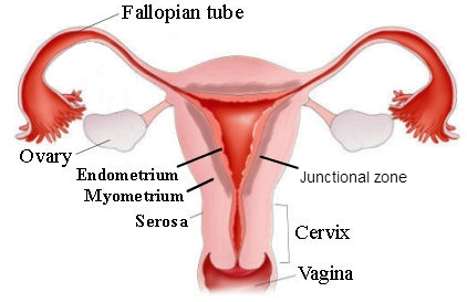
After ovulation, the ovum travels into an oviduct (also called the uterine tube or fallopian tube), one of the two tubes attached to the upper lateral portions of the uterus. These tubes arch above the ovaries and have fingerlike projections (fimbriae) that sweep the released ovum into the oviduct. If fertilization occurs, it usually takes place in the oviduct. The uterus is the organ that nourishes the developing offspring. It is pear-shaped, with an upper rounded fundus, a triangular cavity, and a lower narrow cervix that projects into the vagina. The innermost layer of the uterine wall, the endometrium, has a rich blood supply. It receives the fertilized ovum and becomes part of the placenta during pregnancy. The endometrium is shed during the menstrual period if no fertilization occurs. The muscle layer of the uterine wall is  the myometrium. The vagina is a muscular tube that receives the penis during intercourse, functions as a birth canal, and transports the menstrual flow out of the body.

 ► THE EXTERNAL GENITAL ORGANS:
All of the external female genital organs together are called the vulva. This includes the large outer labia majora and small inner labia minora that enclose the openings of the vagina and the urethra. The clitoris, anterior to the urethral opening, is similar in origin to the penis and responds to sexual stimulation. In both male and female, the region between the thighs, from the external genital organs to the anus, is the perineum. During childbirth, an incision may be made between the vagina and the anus to facilitate birth and prevent the tearing of tissue, a procedure called an episiotomy.

 ► Mammary Gland

The mammary glands, or breasts, are composed mainly of glandular tissue and fat.   (((((( Their purpose is to provide nourishment for the newborn))))). . The milk secreted by the glands is carried in ducts to the nipple. The Menstrual Cycle: Reproductive activity in the female normally begins during puberty with menarche, the first menstrual period indicates that girl is matuar and she is ready to give birth.and from that time mensuration continue at Each month, the menstrual cycle is controlled,  by hormones from the anterior pituitary gland. Follicle stimulating hormone (FSH) begins the cycle by causing the ovum to ripen in the graafian follicle. The follicle secretes estrogen, a hormone that starts development of the endometrium in preparation for the fertilized egg. A second pituitary hormone, luteinizing hormone (LH), triggers ovulation and conversion of the follicle to the corpus luteum. This structure, left behind in the ovary, secretes progesterone and estrogen, which further the growth of the endometrium. If no fertilization occurs, hormone levels decline, and the endometrium sloughs off in the process of menstruation. The average menstrual cycle lasts 28 days, with the first day of menstruation taken as day 1 and ovulation occurring on about day 14. Throughout the cycle, estrogen and progesterone feed back to the pituitary to regulate the production of FSH and LH.     Family planning;  Hormonal methods of birth control act by supplying estrogen and progesterone, which inhibit the pituitary and prevent ovulation, while not interfering with menstruation.  Contraception: Contraception is the use of artificial methods to prevent fertilization of the ovum or its implantation in the uterus. Methods can be used to block sperm penetration of the uterus (condom, diaphragm), prevent implantation (intrauterine device [IUD]), or prevent ovulation (hormonal methods). Surgical sterilization for the male is a vasectomy; for the female, surgical sterilization is a tubal ligation, in which the fallopian tubes are cut and tied on both sides. The preferred method for performing this surgery is through the abdominal wall with a laparoscopes. Unprotective  sexual intercourse is the major deseas causing factors of this system.so be careful. You can read this article in hausa by clicking here .and for any corrections or suggestions call me at or send to junaiduumar2233@gmail.com.           

Sunday, January 13, 2019

(18).RESPIRATORY SYSTEM

Overview :
The main function of the respiratory system is to provide oxygen to body cells for energy metabolism and to eliminate carbon dioxide, a byproduct of metabolism. Because these gases must be carried to and from the cells in the blood, the respiratory system works closely with the cardiovascular system to accomplish gas exchange. Exchange of gases between the atmosphere and the blood takes place in the lungs. A double membrane, the pleura, covers the lungs and lines the thoracic cavity. The outer layer that is attached to the wall of the thoracic cavity is the parietal pleura; the inner layer that is attached to the surface of the lungs is the visceral pleura.
 The very thin, fluid-filled space between the two layers of the pleura is the pleural space.
 Upper Respiratory Passageways: Air is carried to and from the lungs in a series of tubes in which no gas exchange occurs. Air enters through the nose, where it is warmed, filtered, and moistened as it passes over the hair-covered mucous membranes of the nasal cavity. Cilia, microscopic hair-like projections from the cells that line the nose, sweep dirt and foreign material toward the throat for elimination. Material that is eliminated from the respiratory tract by coughing or clearing the throat is called sputum. In the bones of the skull and face near the nose are air-filled cavities lined with mucous membranes that drain into the nasal cavity. These chambers lighten the bones and provide resonance for speech production. Each of these cavities is called a sinus, and they are named specifically for the bones in which they are located, such as the sphenoid, ethmoid, and maxillary sinuses. Together, because they are near the nose, these cavities are referred to as the paranasal sinuses. Receptors for the sense of smell are located within bony side projections of the nasal cavity called turbinate bones or conchae. Inhaled air passes into the throat, or pharynx, where it mixes with air that enters through the mouth and also with food destined for the digestive tract. The pharynx is divided into three regions:
(1) an upper portion, the nasopharynx, behind the nasal cavity;
 (2) a middle portion, the oropharynx, behind the mouth; and
 (3) a lower portion, the laryngeal pharynx, behind the larynx. The palatine tonsils are on either side of the soft palate in the oropharynx; the pharyngeal tonsils, or adenoids, are in the nasopharynx.
 Lower Respiratory Passageways:  and Lungs The pharynx conducts air into the trachea, a tube reinforced with C-shaped rings of cartilage to prevent its collapse (you can feel these rings if you press your fingers gently against the front of your throat). Cilia in the lining of the trachea move impurities up toward the throat, where they can be eliminated by swallowing or by expectoration. At the top of the trachea is the larynx. The larynx is shaped by nine cartilages, the most prominent of which is the thyroid cartilage at the front that forms the “Adam’s apple.” The opening between the vocal cords is the glottis. The small leaf-shaped cartilage at the top of the larynx is called the epiglottis. When one swallows, the epiglottis covers the opening of the larynx and helps to prevent food from entering the respiratory tract.
 The larynx contains the vocal cords, folds of tissue that are important in speech production. Vibrations produced by air passing over the vocal cords form the basis for voice production, although portions of the throat and mouth are needed for proper articulation of speech.
 The trachea is contained in a region known as the mediastinum, which consists of the space between the lungs together with the organs contained in this space. In addition to the trachea, the mediastinum contains the heart, esophagus, large vessels, and other tissues. At its lower end, the trachea divides into a right and a left main stem bronchus that enter the lungs. The right bronchus is shorter and wider; it divides into three secondary bronchi that enter the three lobes of the right lung. The left bronchus divides into two branches that supply the two lobes of the left lung. Further divisions produce an increasing number of smaller tubes that supply air to smaller subdivisions of lung tissue. As the air passageways progress through the lungs, the cartilage in the walls gradually disappears and is replaced by smooth (involuntary) muscle. The smallest of the conducting tubes, the bronchioles, carry air into the microscopic air sacs, the alveoli, through which gases are exchanged between the lungs and the blood. It is through the ultrathin walls of the alveoli and their surrounding capillaries that oxygen diffuses into the blood and carbon dioxide diffuses out of the blood for elimination.

► BREATHING

Air is moved into and out of the lungs by the process of breathing, technically called ventilation. This consists of a steady cycle of inspiration (inhalation) and expiration (exhalation), separated by a period of rest. The cycle begins when the phrenic nerve stimulates the diaphragm to contract and flatten, thus enlarging the chest cavity. The resulting decrease in pressure within the thorax causes air to be pulled into the lungs. The intercostal muscles between the ribs aid in inspiration by pulling the ribs up and out. Muscles of the neck and thorax are used in addition for forceful inhalation. The measure of how easily the lungs expand under pressure is called compliance. Fluid produced within the lung, known as surfactant, aids in compliance by reducing surface tension within the alveoli. Expiration occurs as the breathing muscles relax, the lungs spring back to their original size, and air is forced out. Muscles of the rib cage and abdomen can be called on for forceful exhalation. Breathing is normally regulated unconsciously by centers in the brainstem. These centers adjust the rate and rhythm of breathing according to changes in the composition of the blood, especially the concentration of carbon dioxide.

► GAS TANSPORT :

Oxygen is carried in the blood bound to hemoglobin in red blood cells. The oxygen is released to the cells as needed. Carbon dioxide is carried in several ways but is mostly converted to an acid called carbonic acid. The amount of carbon dioxide that is exhaled is important in regulating the acidity or alkalinity of the blood, based on the amount of carbonic acid that is formed. Dangerous shifts in blood pH can result from too much or too little carbon dioxide being exhaled.to help this system works properly Regular exercise and deep breath is very important.if you like to read this turtorial in hausa, visit my alternated website,at
Www.kimiyyadahausa.blogspot.com.for any question or corrections leave your comment in below box.
Than to all of us.

Earth science

(2).Introduction to earth science